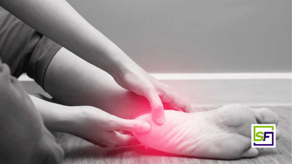
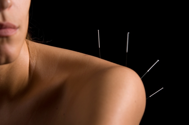
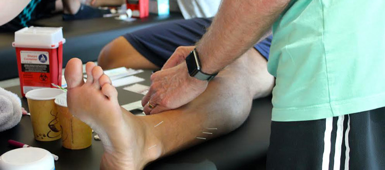
In this article we will discuss the pathophysiology of plantar heel pain and the current research into treating plantar heel pain with dry needling.
Plantar heel pain is a common problem among adults. Each year in the United States, people visit the physician more than 1 million times per year because of plantar heel pain.1 It is estimated that plantar heel pain accounts for 80% of patients with inferior heel pain and affects 1 in 10 people in their lifetime.2 It afflicts athletes and sedentary people alike and affects both males and females equally. Identifying the main cause of the pain can be complicated and multifactorial, and despite its prevalence, the nexus of plantar heel pain is not well understood.3 The most frequent cause of plantar heel pain is plantar fasciitis affecting almost 10% of the population over a lifetime.1 However, plantar heel pain can occur from calcaneal stress fractures or lower extremity radiculopathies as well.2,4 These conditions can lead to severe pain, which causes significant disability and impact the daily lives of those who suffer from these conditions. These conditions are commonly treated with lifestyle modification strategies, non-steroidal anti-inflammatory drugs (NSAIDs), physical rehabilitation, local injections, and potentially even surgery.1,4,5
Pathophysiology of Plantar Heel Pain
Plantar Fasciitis
The posterior tuberosity of the calcaneus has medial and lateral processes. The medial process gives rise to the attachment to the Flexor digitorum brevis, Abductor hallucis, the medial head of quadratus plante, and the plantar fascia’s central band. It is here where many patients experience heel pain. The plantar fascia includes medial, mid, and lateral portions. The most important is the central piece, which attaches proximally has a direct fibrocartilaginous attachment to the calcaneus. The fascia’s triangular shape develops from the medial process of the calcaneal tuberosity and diverges distally at the mid-metatarsal level into five separate strands. These are attached at the forefoot onto the plantar skin, the base of proximal phalanges, and the metatarsophalangeal joints via the collateral ligaments and deep, transverse metatarsal ligaments.1,2,6 It supports the longitudinal arch, and it elongates with rising mechanical loading. Obesity, prolonged standing, flat feet, soleus-gastrocnemius complex dysfunction, and ankle instability can lead to excessive stress on the fascia. Aging leads to a reduction of the elasticity of the plantar fascia. All the factors mentioned above will lead to the plantar fascia’s degeneration rather than inflammation and thickening. 1,2,6 Obesity, pes planus, posterior chain dysfunction, and ankle instability can lead to excessive stress on the fascia.1 Aging leads to a reduction of the elasticity of the plantar fascia. All the factors mentioned above will lead to the degeneration of the plantar fascia rather than inflammation and thickening.1,6
Calcaneal Stress Fractures
A calcaneal stress fracture is a small break in the heel bone, also known as the calcaneus, due to repetitive activity on foot. Normal weight-bearing stress is less than the threshold needed to cause a calcaneal stress fracture. That repetitive activity can cause small amounts of trauma to the bone. Without enough healing time between workouts, it can result in microscopic damage that can progress to a stress fracture.1,2,6 This typically happens with overuse or the start of new activities that include running or bounding. Other relevant factors beyond starting a new exercise routine include increased intensity or duration of previous activities, obesity, inappropriate footwear, and pes cavus foot.1,6 The calcaneus is the second most common location for stress fractures in the foot.1
Entrapment Neuropathies
Tarsal tunnel syndrome is due to the tibial nerve’s entrapment underneath the flexor retinaculum on the ankle’s medial side. It might be idiopathic or secondary to diabetes mellitus and ganglion cysts.1 Also, the medial calcaneal nerve commonly arises from the tibial nerve above the ankle’s flexor retinaculum. Heel skin is innervated by the medial calcaneal nerve, which may present with heel pain if compressed proximally by some pathology. Other entrapment is possible at Baxter’s nerve. Baxter’s nerve is the first branch of the lateral plantar nerve and carries sensory information from the calcaneal periosteum and the long plantar ligament. Baxter’s nerve can be entrapped distally due to tight fascial planes between the abductor hallucis muscle and the quadratus plantae. Another entrapment site is at the anterior aspect of the medial calcaneal tuberosity between the flexor digitorum brevis and quadratus plantae.1,6,7
Dry Needling for Plantar Heel Pain
There were two recent articles published in 2020 that directly relate to dry needling for plantar heel pain. Al-Boloushi et al. executed a randomized control trial that compared two dry needling interventions for plantar heel pain. They attempted to compare the effectiveness of dry needling versus dry needling with electrical stimulation for improving the level of pain, function, and quality of life of patients suffering from plantar heel pain provoked by myofascial trigger points. Subjects were randomized into two groups. One group received dry needling and a stretching protocol, whereas the other group received dry needling with electrical stimulation with a stretching protocol. Measures included the Foot Health Status Questionnaire, a 0–10 numerical rating scale pain visual analog scale (VAS), and a QoL was measured using the EuroQoL-5 dimensions. All measurements were taken at baseline, at 4, 8, 12, 26, and 52 weeks. The results were impressive because the Foot Health Status Questionnaire improved at all time points for the dry needling group and the needling electrolysis group, without significant differences between groups. Pain VAS scores decreased at all time points for both the dry needling and the needling electrolysis group. Additionally, QoL improved at four weeks for both the dry needling and the needling electrolysis group and at 8 and 52 weeks for the needling electrolysis group only. There were significant differences between groups for the QoL at 52 weeks. These effects led them to conclude that both dry needling and dry needling with electric stimulation were sufficient to manage plantar heel pain in the short run. However, additional quality of life changes at 52 weeks in favor of the dry needling with the electric stimulation group.3
This was followed by an article by Wang and colleagues that had a similar approach and similar results. They set out to compare electro-acupuncture and manual acupuncture for plantar heel pain. They randomly assigned participants to receive 12 treatment sessions of electro-acupuncture or manual acupuncture over 4 weeks with a follow-up at 16 and 24 weeks. The primary outcome was the proportion of treatment responders, defined as patients with at least a 50% reduction from baseline in the worst pain intensity experienced during the first steps in the morning after the 4-week treatment, measured using a visual analogue scale (VAS, 0–100; higher scores signify worse pain). Analysis was by intention-to-treat. 54.8% of the electro-acupuncture group compared to 50.0% of the manual acupuncture group achieved a 50% reduction in pain on first steps in the morning after the 4-week treatment. There were no significant between-group differences at 16 weeks and 24 weeks of follow-up. They concluded similar to the previous article that both electro-acupuncture and manual acupuncture appeared to have positive temporal effects, with decreased heel pain and improved plantar function.8
Conclusion
There is now an accumulation of evidence that points to dry needling’s utility in treating plantar heel pain.9–14 These two are just the most recent articles demonstrating that dry needling and electric dry needling interventions are effective. This information informs how we use dry needling for the treatment of plantar heel pain. If you’re interested in learning how Structure & Function Educations’s Pentamodal Method of Dry Needling can be used to treat plantar heel pain, enroll today in a Foundations in Dry Needling for Orthopedic Rehab & Sport Performance course at structureandfunction.net.
References
1. Allam AE, Chang K-V. Plantar Heel Pain. In: StatPearls. StatPearls Publishing; 2020. Accessed January 6, 2021.
http://www.ncbi.nlm.nih.gov/books/NBK499868/
2. Rosenbaum AJ, DiPreta JA, Misener D. Plantar heel pain. Med Clin North Am. 2014;98(2):339-352. doi:10.1016/j.mcna.2013.10.009
3. Al-Boloushi Z, Gómez-Trullén EM, Arian M, Fernández D, Herrero P, Bellosta-López P. Comparing two dry needling interventions for plantar heel pain: a randomised controlled trial. BMJ Open. 2020;10(8):e038033. doi:10.1136/bmjopen-2020-038033
4. Orhurhu V, Urits I, Orman S, Viswanath O, Abd-Elsayed A. A Systematic Review of Radiofrequency Treatment of the Ankle for the Management of Chronic Foot and Ankle Pain. Curr Pain Headache Rep. 2019;23(1):4. doi:10.1007/s11916-019-0745-5
5. Malahias M-A, Cantiller EB, Kadu VV, Müller S. The clinical outcome of endoscopic plantar fascia release: A current concept review. Foot Ankle Surg. 2020;26(1):19-24. doi:10.1016/j.fas.2018.12.006
6. Hossain M, Makwana N. “Not Plantar Fasciitis”: the differential diagnosis and management of heel pain syndrome. Orthopaedics and Trauma. 2011;25(3):198-206. doi:10.1016/j.mporth.2011.02.003
7. Chundru U, Liebeskind A, Seidelmann F, Fogel J, Franklin P, Beltran J. Plantar fasciitis and calcaneal spur formation are associated with abductor digiti minimi atrophy on MRI of the foot. Skeletal Radiol. 2008;37(6):505-510. doi:10.1007/s00256-008-0455-2
8. Wang W, Liu Y, Jiao R, Liu S, Zhao J, Liu Z. Comparison of electro-acupuncture and manual acupuncture for patients with plantar heel pain syndrome: a randomized controlled trial. Acupunct Med. Published online August 18, 2020:096452842094773. doi:10.1177/0964528420947739
9. Wang W, Liu Y, Jiao R, Liu S, Zhao J, Liu Z. Comparison of electro-acupuncture and manual acupuncture for patients with plantar heel pain syndrome: a randomized controlled trial. Acupunct Med. Published online August 18, 2020:096452842094773. doi:10.1177/0964528420947739
10. Al-Boloushi Z, Gómez-Trullén EM, Arian M, Fernández D, Herrero P, Bellosta-López P. Comparing two dry needling interventions for plantar heel pain: a randomised controlled trial. BMJ Open. 2020;10(8):e038033. doi:10.1136/bmjopen-2020-038033
11. Dunning J, Butts R, Henry N, et al. Electrical dry needling as an adjunct to exercise, manual therapy and ultrasound for plantar fasciitis: A multi-center randomized clinical trial. Baur H, ed. PLoS ONE. 2018;13(10):e0205405. doi:10.1371/journal.pone.0205405
12. Ebrahim DAH, Ahmed DGM, Elsayed DE, Sarhan DR. Effect of Electro Acupuncture TENS, Stretching Exercises and Prefabricated Insole in Patients With Plantar Fasciitis. :12.
13. He C, Ma H. Effectiveness of trigger point dry needling for plantar heel pain: a meta-analysis of seven randomized controlled trials. JPR. 2017;Volume 10:1933-1942. doi:10.2147/JPR.S141607
14. Kumnerddee W, Pattapong N. Efficacy of electro-acupuncture in chronic plantar fasciitis: a randomized controlled trial. Am J Chin Med. 2012;40(6):1167-1176. doi:10.1142/S0192415X12500863


