The Importance of Anatomy to the Dry Needling Practice
I was recently asked on a podcast a very interesting question. What are the requisite sets of knowledge and skills to be a safe and effective dry needler? It was a very interesting discussion where we discussed practice history, types of educational backgrounds and skills but my very fundamental and first clear line was a strong knowledge of anatomy and an appreciation for your patient’s anatomy. The importance of anatomy to the dry needling practice is even more important as we start placing needles 3-dimensionally into the body. If you’re interested in this discussion you can check it out on The True North Podcast with Jaime Huestis.
What is Dry Needling?
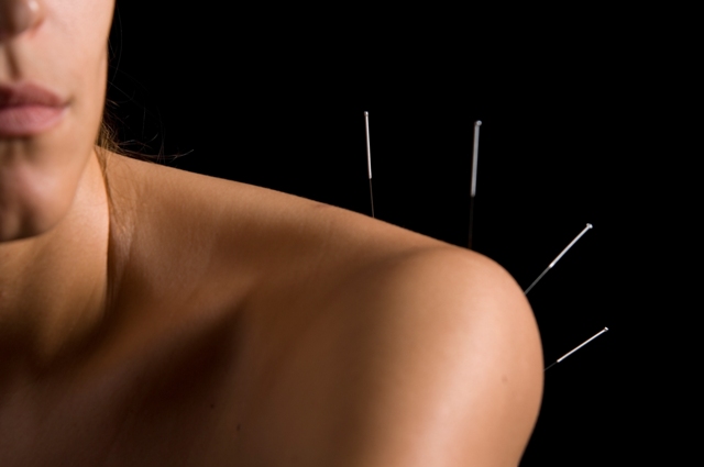
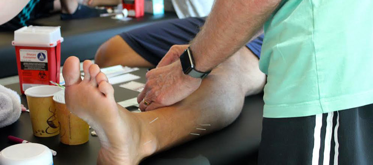
Understand that “dry needling” refers to the insertion of thin monofilament needles without the use of injectate.1–4 Dry-needling is typically used to treat muscles, ligaments, tendons, subcutaneous fascia, scar tissue, peripheral nerves, and neurovascular bundles for the management of a variety of neuromusculoskeletal disorders.1
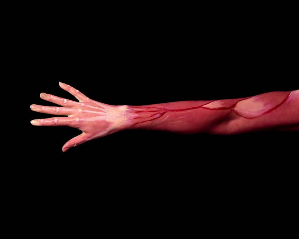
When inserting a needle into the body you have the opportunity to damage structures within the body and can create some major adverse events that are often the focus of discussion such as organ puncture such as pneumothorax, punctured kidneys etc. This is understandable as dry needling involves a needle penetrating the skin and injury to vessels, nerves, spinal cord, internal organs, implanted devices, or infection are possible hazards for patients.5
Several authors have discussed the types of adverse events that can happen when performing dry needling.5–8 In general these paint a picture that dry needling is very safe. Most of the adverse events are very mild and involve a drop of blood, some pain at the needle site and small amounts of bruising. However, some major adverse events can occur.
Brady and colleagues followed the treatments of 39 physiotherapists and 1,463 (19.18%) mild AE’s were reported in 7,629 treatments. They reported no significant adverse events were reported and estimated upper risk rate for significant adverse events to be less than or equal to 0.04%.6 Boyce and colleagues followed this with a larger study where four hundred and twenty physical therapists participated in tracking minor and major adverse events that occurred during 20,464 dry needling treatments. In this study they only had twenty major adverse events reported out of the 20,494 treatments for a rate of less than 0.1%. No associations were noted between the frequency of adverse events and the number of patients treated, practitioner age, level of education, years in practice, level of training or months experience with dry needling.
Importance of Anatomy
Halle and Halle wrote 2 articles that discussed dry needling safety.7,8 They spent two articles discussing that the underlying anatomy across individuals has variability, requiring an in-depth knowledge of anatomy prior to any needle placement. They then provided an overview of pertinent anatomy in the region of the thorax, abdomen, pelvis, back, vasovagal response, informed consent, and other pertinent issues. They concluded that dry needling is an effective adjunct treatment procedure that can be a safe when implementation of the procedure is based on a thorough understanding of the underlying anatomy and the potential risks, with risks coordinated with patients via informed consent.7,8
Many safety studies have been published using cadavers 11–14, diagnostic ultrasound 15–19 and other techniques to help practitioners deepen their understanding of anatomy and dry needling safety. This brings me to a new and interesting article that was just published. Inclusion of Cross-Sectional and Radiological Images for Better Understanding of Musculoskeletal Anatomy and Decreasing the Risk of Adverse Events during Dry Needling in Undergraduate Physiotherapy Students.20 The aim of this study was to analyze the students’ opinion of including cross-sectional and radiological images to traditional methodologies, to evaluate whether these additional resources improve their ability to identify musculoskeletal structures in radiological images and their understanding of neurovascular and visceral structures related with specific muscles to be avoided during invasive procedures.
In general, students appreciated cross-sectional images to be of utility for learning anatomy (81.8%) as well as radiological images (93.9%). All tests showed significant improvements in anatomy knowledge after the inclusion of these complementary resources. They concluded that the implementation of anatomical cross-sectional and radiological images resulted in better understanding of possible risk during invasive procedures.20 Again, demonstrating additional exposure to multiple ways of exploring the body is necessary for learning.
One of the responses we hear about almost every course we teach is that the anatomical review prior to needling had increased the clinician’s understanding and respect of anatomy.
While anatomy knowledge is considered essential by physical therapists, athletic trainers, chiropractors, and other physical medicine providers to be able to effectively apply different treatments based on accurate diagnosis, many haven’t had any anatomic course work in some time. Additionally, they tell me they often see narrow patient populations and “it has been a long time since I treated “insert body area here.”
Although cadaveric dissection is considered a gold standard for teaching anatomy, many clinicians either never had a cadaver dissection experience or it occurred years ago. Universities and body donations programs are reducing exposure to cadaver training and using alternative technologies. This is leading to a feeling that cadaver exploration is obsolete.9 COVID didn’t help this as many programs and donations were refused, and all dissections were stopped in both the educational and research settings.10
How Structure & Function Education Helps You Learn More About Anatomy
I hope that you come away from this understanding that anatomic knowledge and skills are necessary for proper dry needling. At Structure & Function Education, we feel cadaver dissection and exploration are essential to being a good dry needler. As such, we offer a cadaver dissection experience for our students via a 3.5 day, 25-hour lab-based Anatomical Dissection course. In this course practitioners can dissect and explore the human anatomy via fresh-frozen tissue cadavers. This is a full body dissection opportunity. The cadavers are not embalmed, which allows for a more realistic appreciation of tissue quality and fascial connections.
While this class is intended for an improved appreciation for human anatomy as it relates to dry needling safety, we value the relationship of dry needling with other clinical interventions such as manual therapy, therapeutic exercise, and strength and conditioning. This is a great way to increase your anatomic knowledge across the board, which will elevate your clinical practice in infinite ways.
In addition, we also offer every student who enrolls in one of our in-person dry needling courses a free, one year subscription to the 3-D anatomy app called Visual Body. This offers a 3-D anatomic image and the ability to remove layers and appreciate where anatomic structures of concern are most likely located. This is done with the hope that students use this to reflect on the anatomy and plan out correct needle lengths and angles based upon the anatomy of their patient.
As you can tell from our blogs, we take safety seriously in our courses and use methods that assure proper safety in needle application. If you would like to learn dry needling from a company that focuses on patient safety and really focuses on the anatomic knowledge necessary to be a safe needler, enroll in an open course today at www.structureandfunction.net
Stay safe,
Brian Hortz
Brian Hortz, PhD AT
Born in Camden, NJ, Brian received a B.A. in physical education with a concentration in sports medicine from Denison University, a masters degree in sports medicine from Ohio University and his doctoral degree in Exercise Science from Ohio State University. Dr. Hortz is an Instructor and the Director of Research and Education for Structure & Function Education. He has been teaching with Structure & Function Education for several years. Dr. Hortz teaches the Foundations and Advanced courses. In addition to his work with Structure & Function Education, he also has a concierge practice and continues to work one-on-one with athletes to make them well.
References
- Dunning J, Butts R, Mourad F, Young I, Flannagan S, Perreault T. Dry needling: a literature review with implications for clinical practice guidelines. Phys Ther Rev. 2014;19(4):252-265. doi:10.1179/108331913X13844245102034
- Dommerholt J. Dry needling — peripheral and central considerations. J Man Manip Ther. 2011;19(4):223-227. doi:10.1179/106698111X13129729552065
- Perreault T, Flannagan SO, Grubb MT, Grubb R. Mechanisms and dose parameters of electric needle stimulation: clinical considerations – Part I. Acupunct Physiother. Published online 2018:10.
- Perreault T, Grubb MT, Gendron BC, Perez JC, Flannagan SO. Mechanisms and Dose Parameters of Manual Needle Stimulation: Clinical Considerations – Part 2. Acupunct Physiother. Published online 2019:15.
- Boyce D, Wempe H, Campbell C, et al. ADVERSE EVENTS ASSOCIATED WITH THERAPEUTIC DRY NEEDLING. Int J Sports Phys Ther. 2020;15(1):103-113. doi:10.26603/ijspt20200103
- Brady S, McEvoy J, Dommerholt J, Doody C. Adverse events following trigger point dry needling: a prospective survey of chartered physiotherapists. J Man Manip Ther. 2014;22(3):134-140. doi:10.1179/2042618613Y.0000000044
- Halle JS, Halle RJ. PERTINENT DRY NEEDLING CONSIDERATIONS FOR MINIMIZING ADVERSE EFFECTS – PART ONE. Int J Sports Phys Ther. 2016;11(4):651-662.
- Halle JS, Halle RJ. PERTINENT DRY NEEDLING CONSIDERATIONS FOR MINIMIZING ADVERSE EFFECTS – PART TWO. Int J Sports Phys Ther. 2016;11(5):810-819.
- Memon I. Cadaver Dissection Is Obsolete in Medical Training! A Misinterpreted Notion. Med Princ Pract. 2018;27(3):201-210. doi:10.1159/000488320
- Manzanares-Céspedes MC, Dalmau-Pastor M, Simon de Blas C, Vázquez-Osorio MT. Body Donation, Teaching, and Research in Dissection Rooms in Spain in Times of Covid-19. Anat Sci Educ. 2021;14(5):562-571. doi:10.1002/ase.2093
- Hannah MC, Cope J, Palermo A, Smith W, Wacker V. Comparison of two angles of approach for trigger point dry needling of the lumbar multifidus in human donors (cadavers). Man Ther. 2016;26:160-164. doi:10.1016/j.math.2016.08.008
- Mesa-Jiménez JA, Fernández-de-las-Peñas C, Koppenhaver SL, Sánchez-Gutiérrez J, Arias-Buría JL. Cadaveric and in vivo validation of needle placement in the medial pterygoid muscle. Musculoskelet Sci Pract. 2020;49:102197. doi:10.1016/j.msksp.2020.102197
- Fernández-de-las-Peñas C, Mesa-Jiménez JA, Lopez-Davis A, Koppenhaver SL, Arias-Buría JL. Cadaveric and ultrasonographic validation of needling placement in the obliquus capitis inferior muscle. Musculoskelet Sci Pract. Published online October 2019:102075. doi:10.1016/j.msksp.2019.102075
- Kearns G, Gilbert KK, Allen B, et al. Accuracy and safety of dry needle placement in the piriformis muscle in cadavers. J Man Manip Ther. 2018;26(2):89-96. doi:10.1080/10669817.2017.1346745
- Wang-Price S, Zafereo J, Couch Z, Brizzolara K, Heins T, Smith L. Short-term effects of two deep dry needling techniques on pressure pain thresholds and electromyographic amplitude of the lumbosacral multifidus in patients with low back pain – a randomized clinical trial. J Man Manip Ther. Published online January 17, 2020:1-12. doi:10.1080/10669817.2020.1714165
- Wang-Price SS, Etibo KN, Short AP, Brizzolara KJ, Zafereo JA. Validity and reliability of dry needle placement in the deep lumbar multifidus muscle using ultrasound imaging: an in-vivo study. J Man Manip Ther. Published online March 22, 2022:1-8. doi:10.1080/10669817.2022.2051239
- Albin SR, Hoffman LR, MacDonald CW, et al. Ultrasonographic Validation for Needle Placement in the Tibialis Posterior Muscle. Int J Sports Phys Ther. 16(6):1541-1547. doi:10.26603/001c.29854
- Bagcier F, Yurdakul OV. A safer way of dry needling therapy for gastrocnemius muscles: ultrasound guidance. Med Ultrason. 2020;22(4):495. doi:10.11152/mu-2840
- Ball A, Perreault T, Fernández-de-las-Peñas C, Agnone M, Spennato J. Ultrasound Confirmation of the Multiple Loci Hypothesis of the Myofascial Trigger Point and the Diagnostic Importance of Specificity in the Elicitation of the Local Twitch Response. Diagnostics. 2022;12(2):321. doi:10.3390/diagnostics12020321
- Valera-Calero JA, Navarro-Santana MJ, Fernández-de-las-Peñas C, et al. Inclusion of Cross-Sectional and Radiological Images for Better Understanding of Musculoskeletal Anatomy and Decreasing the Risk of Adverse Events during Dry Needling in Undergraduate Physiotherapy Students. Anat Sci Educ. n/a(n/a). doi:10.1002/ase.2251